
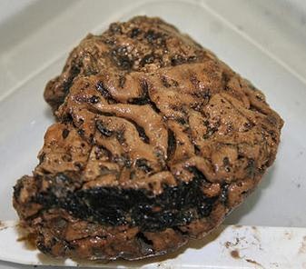
In 2008, in Heslington, a suburban village near modern-day York, England, a brain was found which remained intact for 2600 years. Immunoelectron microscopy confirmed the preservation of neuro-cytoarchitecture in the ancient brain, which appeared shrunken and compact compared to a modern brain. After more than 10 years of research, scientists have found why did the brain survived for so long. Interestingly, protein aggregation typically comes along with brain diseases like Alzheimer’s and Parkinson’s, although the researchers didn’t find evidence of these conditions in the ancient brain. This means the aggregation is not an amyloid plaque or other structures that form after the destruction of the neurofilaments when neuron bursts in the Alzheimers brain. For this particular reason, I got interested in this topic, it is so interesting that there is a possibility of true preservation.
When a person dies a process called autolysis kickstarts, which causes tissues and organs to break down. Human proteins do not survive at ambient temperature, for long periods, the secondary structures begin to unfold and then the chain falls apart. The human brain rapidly dissolves after death due to auto-proteolysis (Auto-proteolysis is the splitting of a protein or peptide into smaller molecules which is catalyzed by the enzymic activity of the protein or peptide itself) and putrefaction (Putrefaction involves the decomposition of proteins, breakdown of cohesiveness between the tissues, and liquefaction of most organs. The body is decomposed by the action of putrefying bacteria and fungi which releases certain gases that infiltrate and deteriorate the body tissues and organs). The brain is 80 percent water and one of the first organs to go down, during degradation, water comes out and the structures are filled with water. Within five to 10 years, the brain tissue is typically totally degraded.
The Heslington brain actually preserved itself. When researchers took a closer look, they discovered that certain proteins, which support the structure of nerve and brain cells like neurons and astrocytes, were clustered close together in the brain tissue. The protein aggregation kept the brain tissue stable over time, helping slow down the natural decomposition process. Immunoassays on micro-dissected brain tissue homogenates revealed the preservation of the known protein topography for grey and white matter for type III (glial fibrillary acidic protein, GFAP) and IV (neurofilaments, Nfs) IFs. Mass spectrometry data could be matched to a number of peptide sequences, notably for GFAP and Nfs. Glial fibrillary acidic protein (GFAP) is a protein that is encoded by the GFAP gene in humans. It is a type III intermediate filament (IF) protein that is expressed by numerous cell types of the central nervous system (CNS), including astrocytes and ependymal cells during development. To form networks, the initial GFAP dimers combine to make staggered tetramers, which are the basic subunits of an intermediate filament. Since rod domains alone in vitro do not form filaments, the non-helical head and tail domains are necessary for filament formation.[17] The head and tail regions have greater variability of sequence and structure. In spite of this increased variability, the head of GFAP contains two conserved arginines and an aromatic residue that have been shown to be required for proper assembly. Neurofilaments (NF) are classed as type IV intermediate filaments found in the cytoplasm of neurons. They are protein polymers measuring 10 nm in diameter and many micrometers in length. Together with microtubules (~25 nm) and microfilaments (7 nm), they form the neuronal cytoskeleton. They are believed to function primarily to provide structural support for axons and to regulate axon diameter, which influences nerve conduction velocity. The proteins that form neurofilaments are members of the intermediate filament protein family, which is divided into six types based on their gene organization and protein structure.
Surprisingly, the researchers also found that the Heslington neural proteins were more stable than modern-day brains. Protein stability is an indirect signal of how well a protein is functioning, the researchers explain. In this case, scientists aren’t sure what sparked this critical clumping process but speculate that it may relate to where and how the skull was buried. Preserved immunogenicity of the prehistoric human brain proteins was demonstrated by antibody gen- eration (GFAP, Nfs, myelin basic protein). Unlike brain proteins, DNA was of poor quality preventing reliable sequencing. These long-term data from a unique ancient human brain demonstrate that aggregate formation permits for the preservation of brain proteins for millennia.
Source:
https://www.inverse.com/article/62194-how-an-ancient-human-brain-survived-2600-years and Wikipedia
